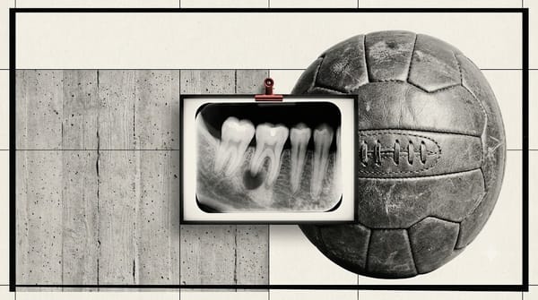
Interproximal Caries on a Bitewing: Why the X-Ray Makes It Look Smaller Than It Is
Interproximal caries hides under the contact point and looks smaller on a bitewing than it really is. Here's how to read the dark triangle and when to treat.

Interproximal caries is tooth decay between two teeth, right under the spot where they touch. On a bitewing X-ray it shows up as a small dark triangle. Here's the catch: the X-ray makes the lesion look smaller than it truly is. A spot that looks stuck in enamel is often already in dentin.
In other words — the X-ray is lying to you. Here's how to read it anyway.
"Students treat the bitewing like a ruler. It isn't — it's a hint, and it always rounds down." — Dr. WS (@dr.WSdent)
What is interproximal caries?
"Interproximal" just means between two teeth. (You'll also hear "proximal" or "approximal" — same thing.) The decay starts just below the contact point: the tiny spot where two teeth press together.
That spot is a trap. Your toothbrush can't reach it. Floss often skips it. Plaque sits there and feeds. So decay starts in the one place that's hardest to clean and hardest to see.
You usually can't see it in the mouth. You usually can't feel it. No pain. No hole you can spot. A 22-year-old can come in for a routine check with zero symptoms — and the bitewing still finds decay hiding under the contact. That's not unusual. That's the whole reason we take the film.
Why do dentists use bitewing X-rays to find it?
Because your eyes miss it. A plain look-and-poke exam catches far less proximal decay than a bitewing does — for early lesions between the teeth, looking alone misses most of them.
The bitewing is the best everyday tool we have for these surfaces. It lines the back teeth up so you can see into the gaps between them.
But — and this matters — the bitewing is not perfect. In one study that checked films against the actual tissue under a microscope, bitewings caught only about 40% of lesions that were still in enamel. They did a little better once decay reached dentin, around 55–60%. The good news: when a film clearly says "yes, decay," it's usually right (specificity was about 88%). The bad news: a clean-looking film does not mean clean teeth.
Translation: the film finds more than your eyes do. It still misses early stuff. So treat "nothing on the X-ray" with humility.
Why does the cavity look smaller on the X-ray than it really is?
This is the big one. Three reasons.
1. The film is late. A proximal lesion usually doesn't even show up on a radiograph until about 30–40% of the mineral is already gone. By the time you see a shadow, real, irreversible damage has started.
2. You're looking through sound enamel. The lesion is a 3-D cone, but the X-ray flattens it into a 2-D picture. Healthy enamel in front of and behind the decay piles on top of it in the image and hides part of it. The deeper the lesion goes, the more sound enamel sits over it — so the deeper it gets, the more the film hides.
3. Measured depth runs short. When researchers compared depth on the film to the true depth under a microscope, the film came up short. In one study of deep lesions, the dentin looked about 20% thicker on the X-ray than it really was — which makes the decay look farther from the nerve than it actually is.
So when you catch yourself thinking "eh, it's barely touching dentin" — assume it's already inside. The shadow is the floor of the damage, not the ceiling.
How deep is it, and how do you read the dark triangle?
A proximal lesion has a shape: a dark triangle, wide at the outer surface, with its tip pointed at the pulp (the nerve). The deeper that tip, the worse the story.
Dentists grade the depth in steps:
- E1 — outer half of enamel
- E2 — inner half of enamel, up to the junction (the DEJ, where enamel meets dentin)
- D1 — outer third of dentin (just crossed the junction)
- D2 — middle third of dentin
- D3 — inner third of dentin (close to the nerve)
Why grade it? Because depth predicts whether the surface has actually broken open — cavitated — and cavitation is what usually decides treatment.
Here's the rough map from the research:
- Still in enamel (E1–E2): usually not cavitated. Only about a quarter to a third have a true hole.
- Outer dentin (D1): the odds flip. Most are cavitated — studies put it around 80–85%+.
- Deep dentin (D2–D3): nearly all are cavitated, and you're getting close to the nerve (often 96–100%).
Now remember reason #1 from above: because the film underestimates, a lesion that looks like E2 may already behave like D1. When in doubt, lean deeper.
Should you drill it or watch it?
Old habit: see decay, drill it. The modern, evidence-based habit: match the treatment to the depth and to whether it's cavitated.
- Enamel only, not cavitated (E1–E2): don't drill. This is the stage you can actually reverse. Clean it up, fluoride, fix the diet and home care, and monitor it. Remineralize, don't restore.
- Into dentin / cavitated (D1 and deeper): restore it. Once it's a real hole into dentin, it won't heal on its own.
The rule in one line: Enamel = watch. Dentin = treat.
One caveat: high-caries-risk patients cavitate sooner, so for them the threshold to treat can shift earlier. Read the patient, not just the pixel.
How do you get fast at spotting these on X-rays?
Pattern recognition. On a busy clinic day you don't reason your way to a diagnosis — you recognize it. And you only recognize what you've already seen a hundred times.
That's the whole point of reps: see a case, call the depth, check yourself, repeat. The dark triangle, the tip, the junction, the "is it cavitated?" call — they stop being scary once they're familiar.
Want the cheat-sheet version of everything above — the depth ladder, the cavitation odds, and the watch-vs-treat rule on a single page? Grab the free Bitewing Caries Cheat Sheet at papilla.io. And if you want the reps, Papilla turns real X-ray cases into a game so you can practice the call until it's automatic.
Frequently asked questions
Can you have a cavity with no pain and nothing visible? Yes. Interproximal decay sits under the contact point, where you can't see or feel it. Many of these are found only on a bitewing X-ray, in people with zero symptoms. Pain usually shows up late, once the decay is deep.
Do X-rays show how deep a cavity is? Partly — and they tend to underestimate it. A proximal lesion usually doesn't appear until about 30–40% of the mineral is lost, and overlapping healthy enamel hides part of the depth. The true lesion is almost always deeper than the film suggests.
What's the difference between E2 and D1 on a bitewing? E2 means the decay is still in enamel, in the inner half, up to the enamel–dentin junction. D1 means it has crossed into the outer third of dentin. The jump matters: E2 lesions are usually not cavitated, while most D1 lesions are.
Should every cavity between teeth be drilled? No. Decay that's still in enamel and not cavitated can often be remineralized and monitored instead of drilled. Drilling is for lesions that have reached dentin or broken the surface. The simple rule: enamel = watch, dentin = treat.
Can AI detect interproximal caries on X-rays? Increasingly, yes. A 2024 review of 21 studies found AI reached about 94% sensitivity and 91% specificity for spotting these lesions on bitewings. But AI is a second opinion, not a substitute — you still need to know how to read the film yourself.
About the author
Dr. WS (@dr.WSdent) is a dentist who now designs and builds learning tools. He got tired of watching students freeze at their first bitewing, so he started breaking real X-ray cases down in plain language. He's the maker behind Papilla, a game-style app that teaches dental X-ray reading one case at a time.
Sources
- Comparative diagnostic accuracy of VistaCam IX Proxi and bitewing radiography for interproximal caries (histology as gold standard) — bitewing sensitivity 40% (enamel), 54.5% (outer dentin), 58.8% (inner dentin); specificity 87.7%. https://www.ncbi.nlm.nih.gov/pmc/articles/PMC10749443/
- Accuracy of radiographic examination in determining depth of approximal lesions (review citing Wenzel; Ratledge) — lesions become radiographically visible only after ~30–40% mineral loss; ~85% of outer-third-dentin lesions are cavitated. http://revodonto.bvsalud.org/scielo.php?script=sci_arttext&pid=S1981-86372012000400008
- Dental Caries (textbook chapter, Pocket Dentistry) — the radiographic image underestimates lesion depth; the triangular lesion is hidden by overlying sound enamel; ~50% of inner-enamel-or-deeper and ~70% of outer-dentin-or-deeper lesions are cavitated. https://pocketdentistry.com/3-dental-caries-2/
- Detection, Activity Assessment and Diagnosis of Dental Caries Lesions (Pocket Dentistry) — radiographs underestimate true histological depth; Pitts & Rimmer threshold (enamel-confined = non-surgical; inner-half dentin = likely cavitated, surgical). https://pocketdentistry.com/detection-activity-assessment-and-diagnosis-of-dental-caries-lesions/
- Clinical cavitation vs radiographic lesion depth (Indian population), Caries/BMC — enamel lesions cavitated 25.6–38.3%; outer dentin 83.3–100%; inner dentin 96.4–100%. https://pubmed.ncbi.nlm.nih.gov/25220524/
- Measurement reliability of remaining dentin thickness below deep lesions — radiographs underestimated remaining dentin thickness by ~20% vs histologic macrophotographs. https://www.ncbi.nlm.nih.gov/pmc/articles/PMC5968158/
- Depth assessment of approximal lesions (DIFOTI vs periapical) — cavitation likelihood and treatment thresholds; lesions extending >halfway to the pulp are most likely cavitated. https://pmc.ncbi.nlm.nih.gov/articles/PMC7191380/
- Diagnostic accuracy of AI for approximal caries on bitewings: systematic review & meta-analysis (21 studies) — pooled sensitivity 0.94, specificity 0.91. https://www.sciencedirect.com/science/article/pii/S030057122400558X
Educational content for dental and hygiene students — not patient-specific diagnostic advice.